
Endoscopic Congenital Heart Defect Surgery
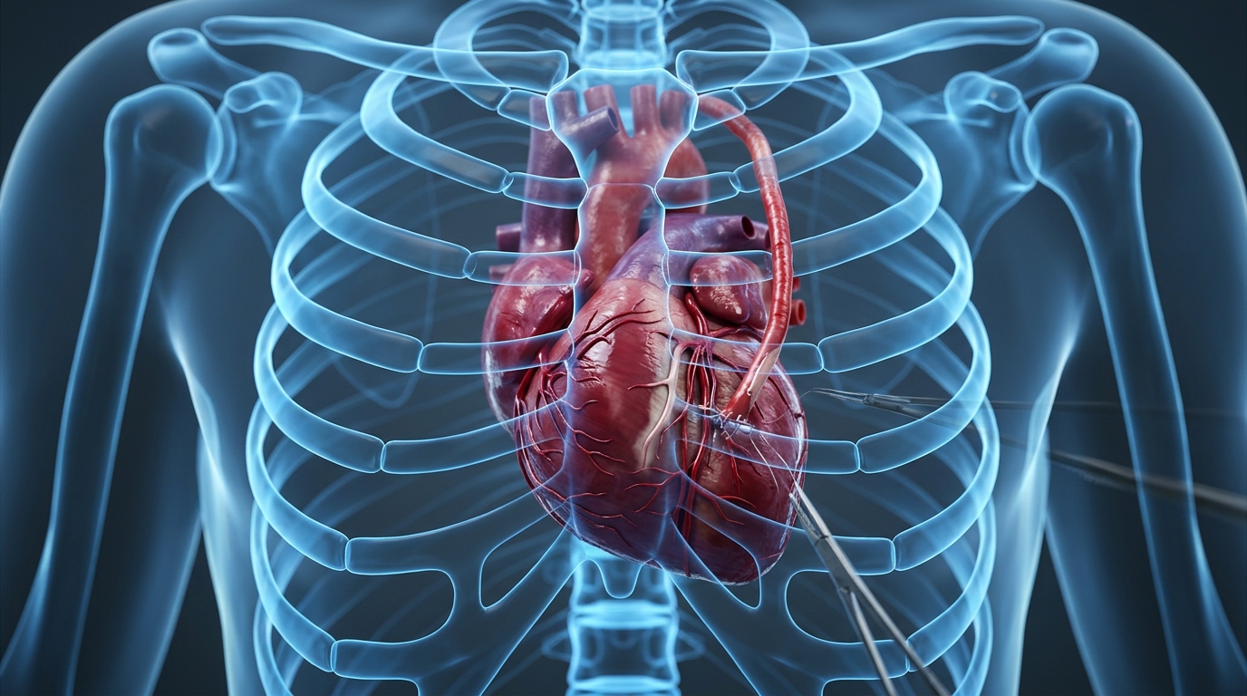
Endoscopic Congenital Heart Surgery (ASD / VSD) performed through an advanced minimally invasive or endoscopic approach without cutting the breastbone, delivering maximum clinical safety and rapid functional recovery.
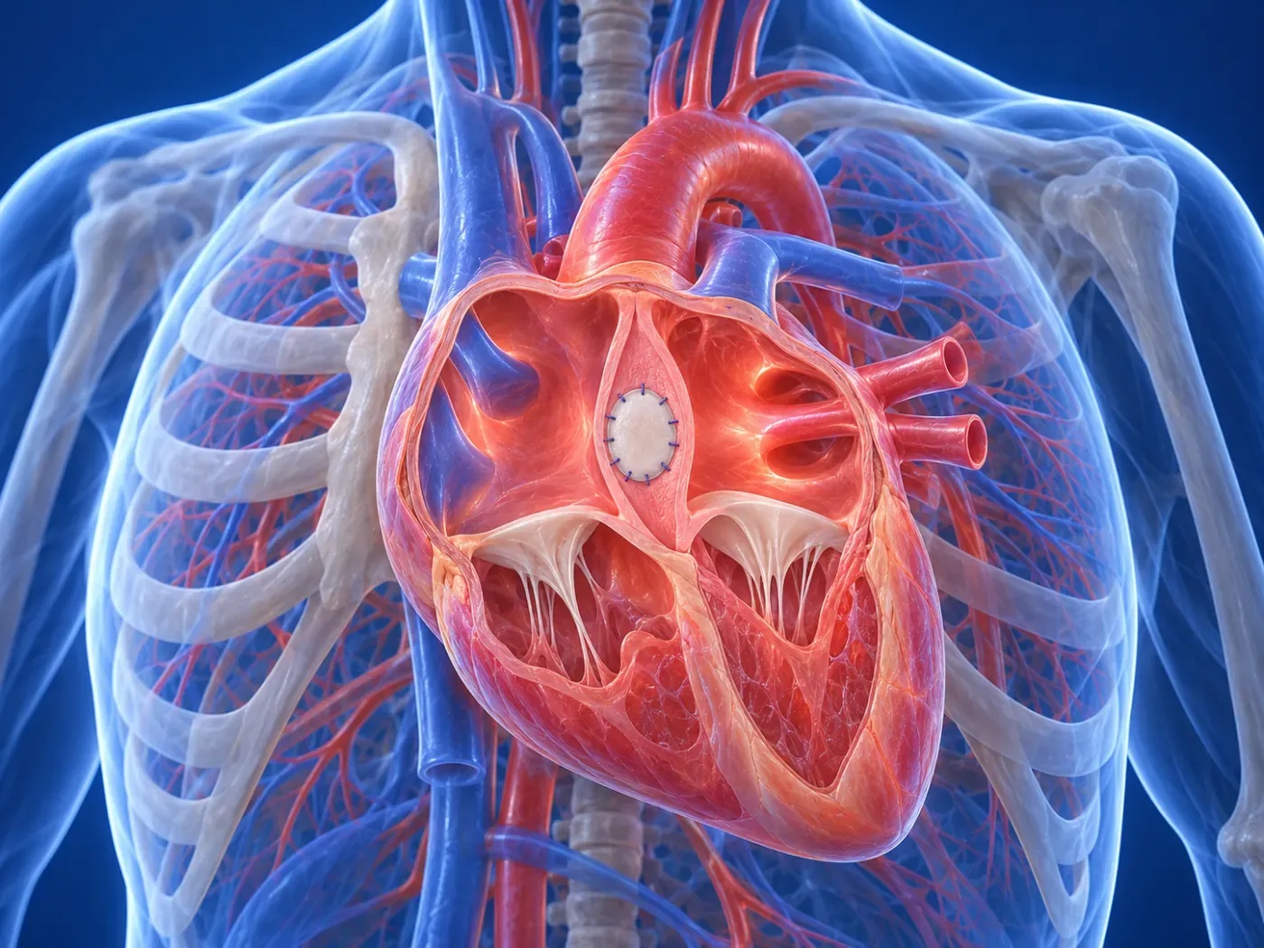
Endoscopic Congenital Heart Surgery (ASD / VSD) performed through an advanced minimally invasive or endoscopic approach without cutting the breastbone, delivering maximum clinical safety and rapid functional recovery.
Clinical Diagnosis & Advanced Indications in Endoscopic Congenital Heart Surgery (ASD / VSD)
Modern diagnostics including 3D imaging, CT angiography, and precision hemodynamic evaluation ensure that patients with Endoscopic Congenital Heart Surgery (ASD / VSD) are treated before irreversible organ or myocardial damage occurs.
Patients receive a comprehensive anatomical assessment allowing our surgical team to plan a tailored procedure that spares the thoracic skeleton entirely.
Surgical Technique & High-Precision Technology
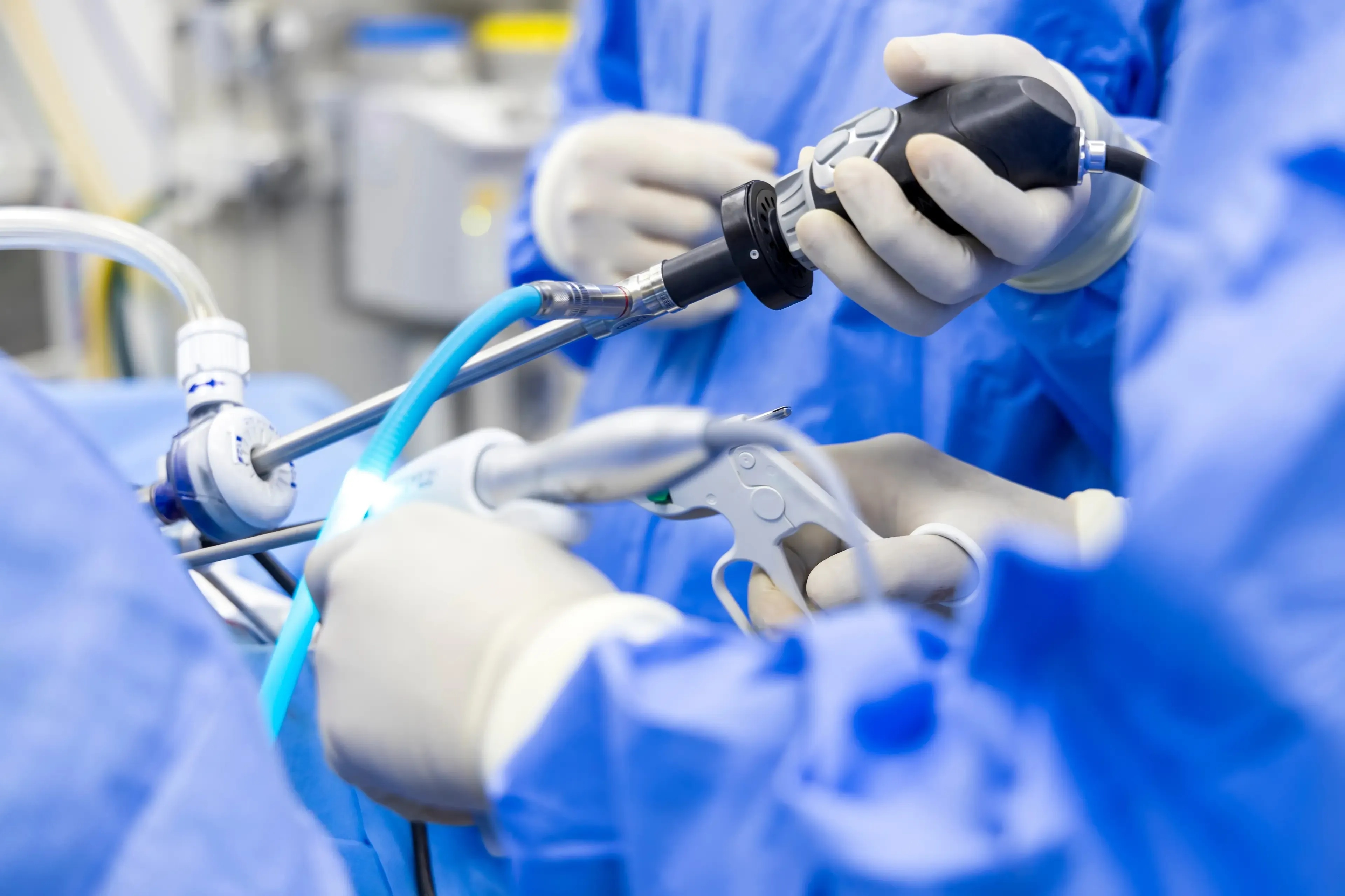
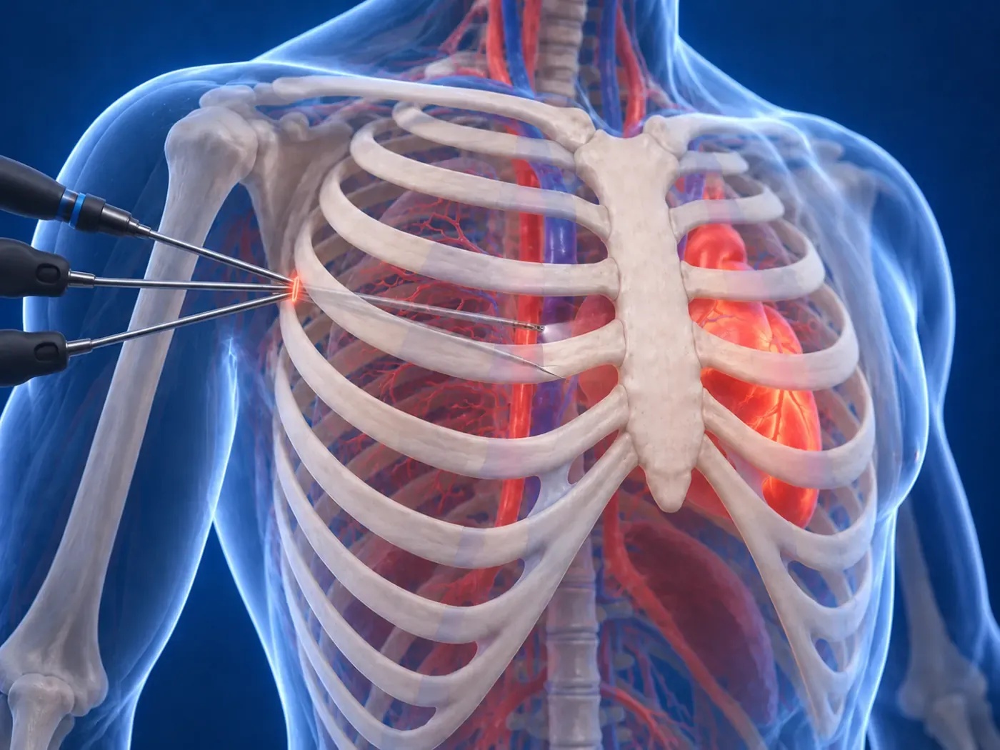
Utilizing specialized micro-instruments, high-definition 3D camera systems, and advanced tissue-preserving methods, the procedure is carried out through small incisions with millimeter accuracy.
Every surgical step is optimized to reduce cross-clamp time, blood trauma, and inflammatory activation, protecting vital organ perfusion.
Key Clinical Advantages Over Traditional Open Surgery
Eliminating full sternotomy transforms the recovery pathway and drastically lowers complication rates:
- The breastbone remains completely intact; no risk of sternal instability or deep bone infection.
- Minimal blood loss and negligible need for blood transfusions.
- Dramatically reduced postoperative pain and zero requirement for rigid chest braces.
- Patients are free to lie in comfortable sleeping positions without restriction.
- Short intensive care stay (24h) and early discharge home within 3 to 4 days.
- Rapid return to work, driving, and active lifestyle in just 10 to 14 days.
Recovery Period, Follow-Up & Consultation
Patients ambulate on the first postoperative day with excellent pulmonary function and minimal pain.
Contact Dr. Taha Özkara with your medical reports and imaging for an in-depth second opinion consultation.
FREQUENTLY ASKED
Frequently Asked Questions
Expert answers to the most common questions regarding minimally invasive heart surgery, recovery timelines, and surgical procedures.
TREATMENT TIMELINE
Your 4-Step Surgical Journey
Tetkiklerinizin incelenmesinden ameliyat sonrası aktif yaşama dönüşünüze kadar geçen 4 aşamalı konforlu ve güvenli cerrahi yolculuğunuz.
Preliminary Review
Comprehensive analysis of your Echo, Angiography, and CT scans.
Tailored Plan
Individual surgical strategy without sternotomy.
Endoscopic Surgery
High-precision closed operation with 3D camera guidance.
Swift Recovery
Discharge in 3-4 days and fast return to normal life.
OUR TREATMENT AREAS
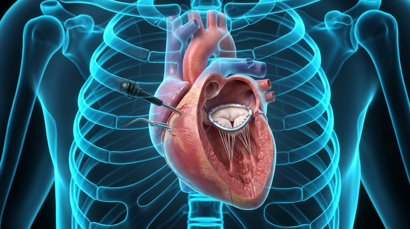
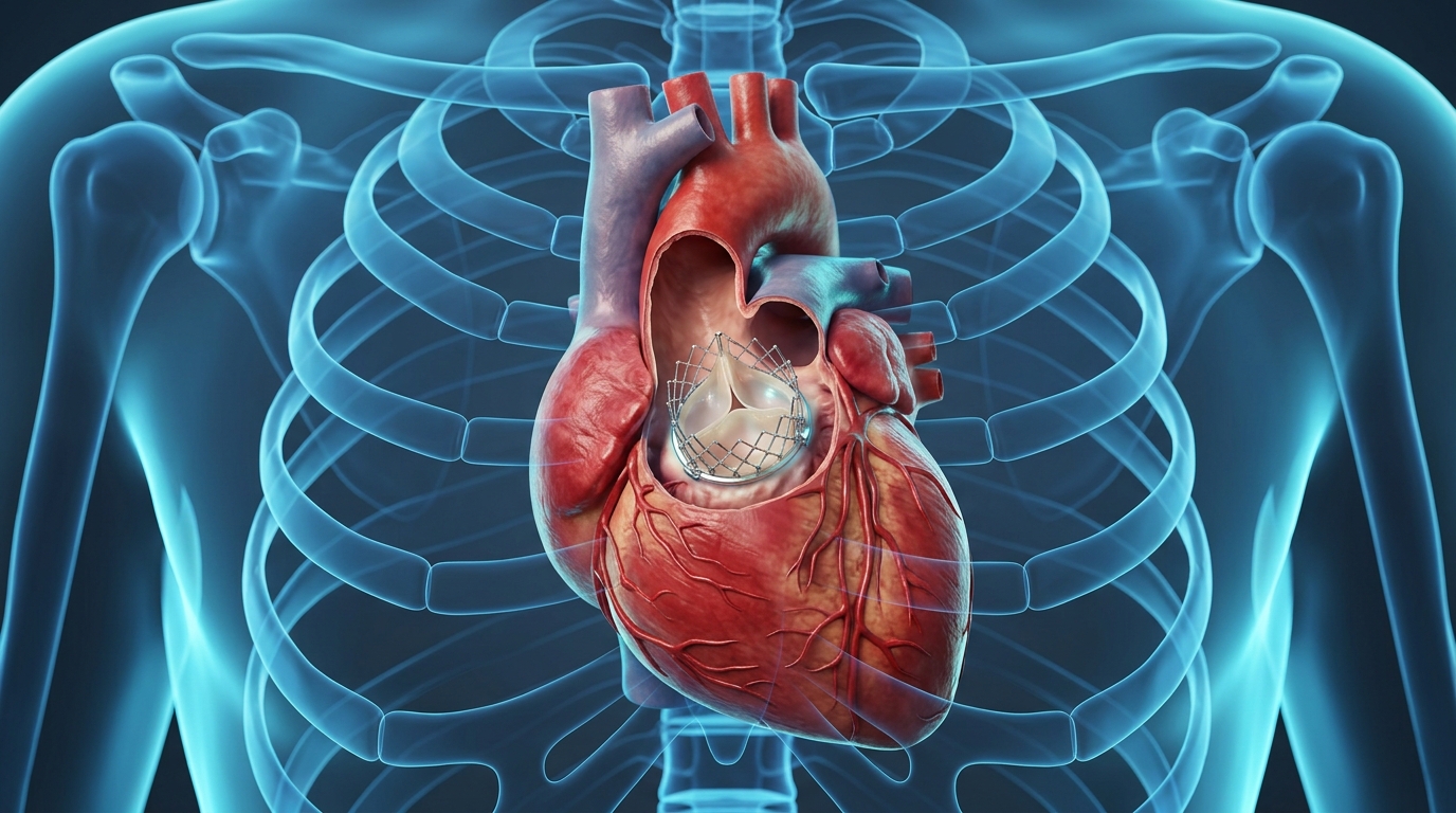
Minimally Invasive and Endoscopic Heart Surgery
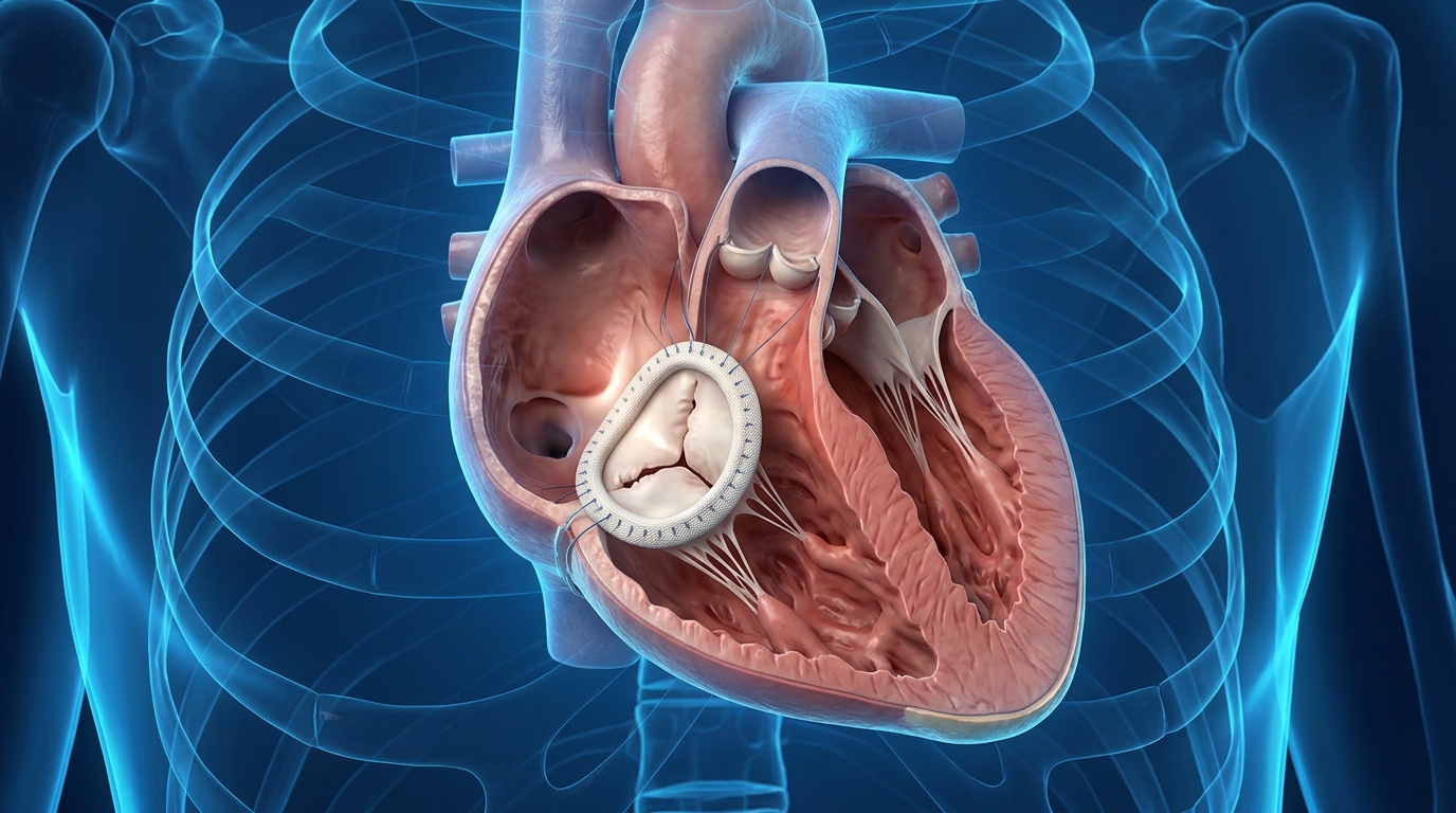
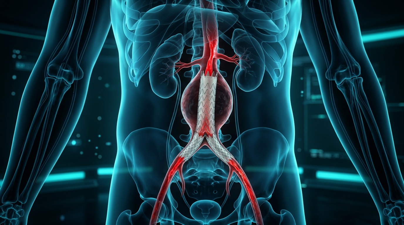
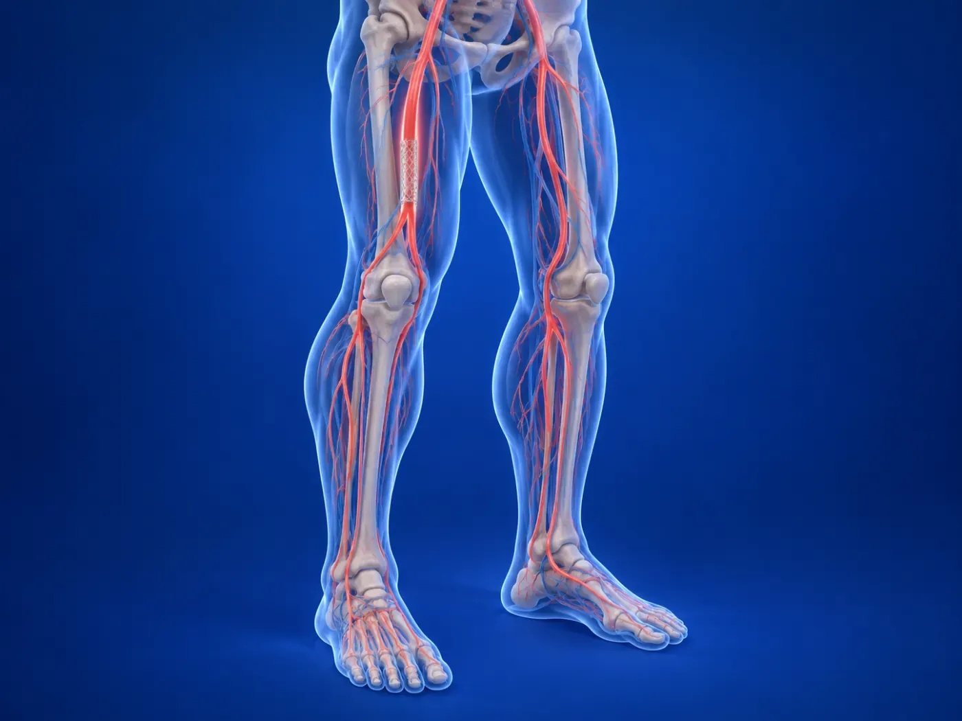
Innovative cardiovascular surgical procedures performed through small incisions with high-definition endoscopic camera guidance without cutting the breastbone.

EXPERT OPINION & APPOINTMENT
Schedule a Medical Consultation
Book your appointment for minimally invasive heart surgery, valve repair, and second opinion consultation.