
Endoscopic Mitral Valve Surgery
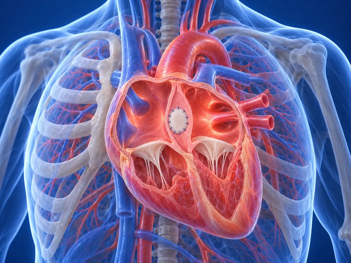
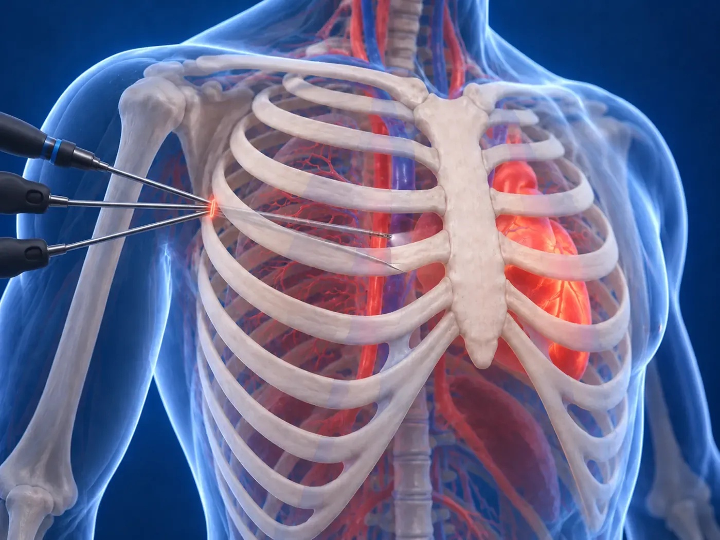
The mitral valve regulates oxygenated blood flow between the heart's left atrium and left ventricle. In modern cardiac surgery, endoscopic mitral valve repair is performed through a small aesthetic incision under the right breast using 3D high-resolution camera guidance without cutting the breastbone, primarily focusing on preserving the patient's own native valve.
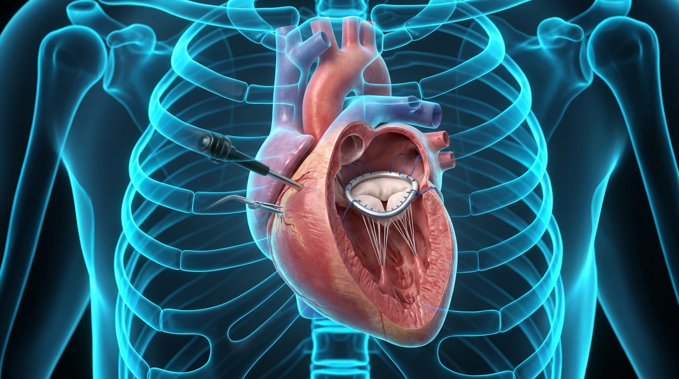
The mitral valve regulates oxygenated blood flow between the heart's left atrium and left ventricle. In modern cardiac surgery, endoscopic mitral valve repair is performed through a small aesthetic incision under the right breast using 3D high-resolution camera guidance without cutting the breastbone, primarily focusing on preserving the patient's own native valve.
Mitral Valve Pathologies and the Endoscopic Approach
Early surgical intervention in mitral valve regurgitation (leakage) or stenosis prevents irreversible left ventricular enlargement and heart failure. Delaying surgery carries severe risks of chronic atrial fibrillation, pulmonary hypertension, and thromboembolism.
Preoperative 3D Transesophageal Echocardiography (3D-TEE) precisely maps leaflet prolapse across P1, P2, P3 or anterior scallops A1, A2, A3, allowing customized surgical repair planning without open sternotomy.
Surgical Technique: Native Valve Repair (Valvuloplasty) & Annuloplasty
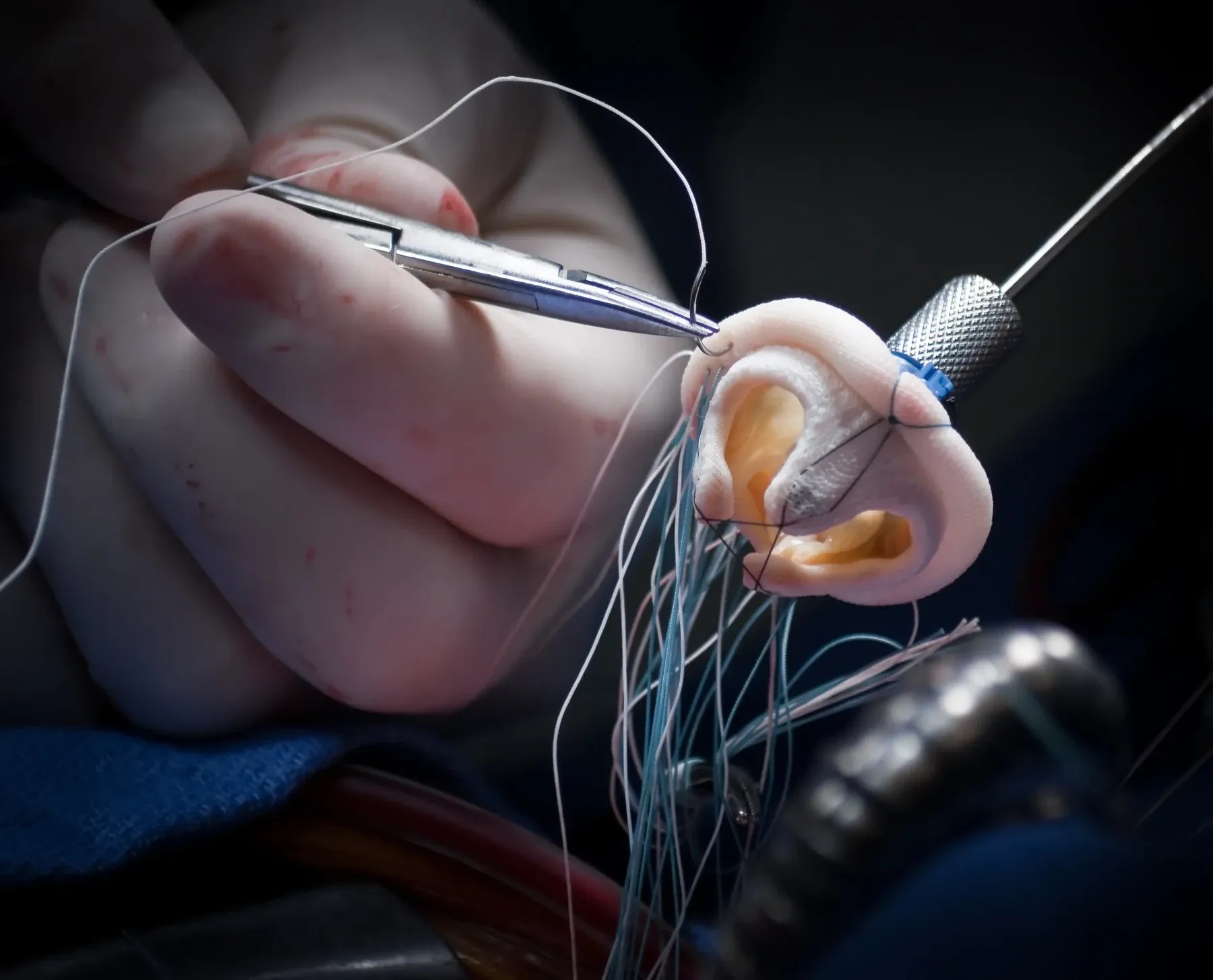
Dr. Taha Özkara prioritizes repairing the patient's native valve over prosthetic replacement whenever possible. Using artificial Gore-Tex neochordae (loops) and triangular/quadrangular resection, normal leaflet coaptation is restored.
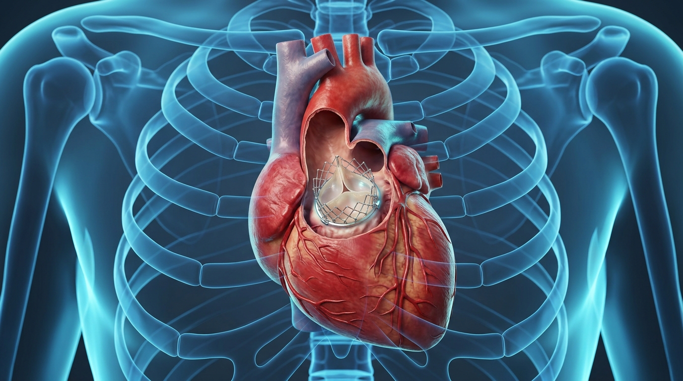
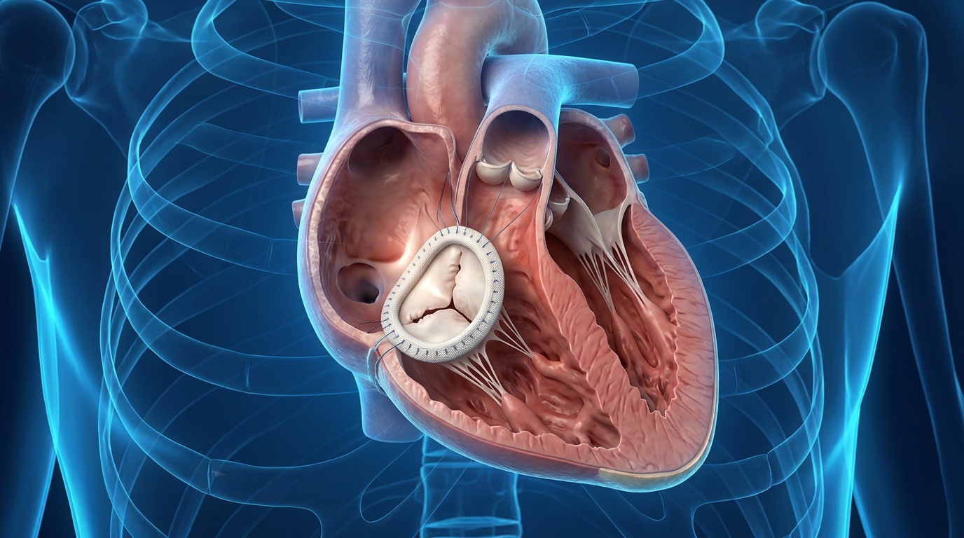
To guarantee lifelong durability, a titanium annuloplasty ring is implanted around the mitral annulus. In severe extensive calcification where repair is unfeasible, biological or mechanical valve replacement is carried out with equal endoscopic safety.
Clinical Advantages of Native Valve Preservation and Closed Surgery
Repairing rather than replacing the valve preserves left ventricular geometry and contractile function, offering superior survival rates and clinical freedom:
- No sternotomy: full thoracic bone stability and respiratory function are preserved.
- Hidden small aesthetic incision under the right breast crease; scar is virtually unnoticeable.
- High-definition 3D endoscopic optics provide millimeter-level surgical precision.
- Preserving the native valve eliminates the requirement for lifelong blood-thinner (warfarin) therapy.
- Significantly lower risks of surgical bleeding and wound infection compared to classical open surgery.
- Discharge in 3-4 days and rapid return to driving, active professional life, and sports in 10-14 days.
Recovery Period, Return to Daily Life, and Patient Assessment
Patients spend only 24 hours in the intensive care unit and are mobilized on postoperative day one. Because the chest bone is intact, sleeping on the back is not mandatory, and patients can sleep on their side comfortably.
All patients with mitral valve insufficiency or stenosis can be evaluated for endoscopic surgery. Contact our surgical team with your echocardiography report for a specialized second opinion.
FREQUENTLY ASKED
Frequently Asked Questions
Expert answers to the most common questions regarding minimally invasive heart surgery, recovery timelines, and surgical procedures.
TREATMENT TIMELINE
Your 4-Step Surgical Journey
Tetkiklerinizin incelenmesinden ameliyat sonrası aktif yaşama dönüşünüze kadar geçen 4 aşamalı konforlu ve güvenli cerrahi yolculuğunuz.
Preliminary Review
Comprehensive analysis of your Echo, Angiography, and CT scans.
Tailored Plan
Individual surgical strategy without sternotomy.
Endoscopic Surgery
High-precision closed operation with 3D camera guidance.
Swift Recovery
Discharge in 3-4 days and fast return to normal life.
OUR TREATMENT AREAS
Minimally Invasive and Endoscopic Heart Surgery
Innovative cardiovascular surgical procedures performed through small incisions with high-definition endoscopic camera guidance without cutting the breastbone.



EXPERT OPINION & APPOINTMENT
Schedule a Medical Consultation
Book your appointment for minimally invasive heart surgery, valve repair, and second opinion consultation.