
Endoscopic Coronary Bypass Surgery
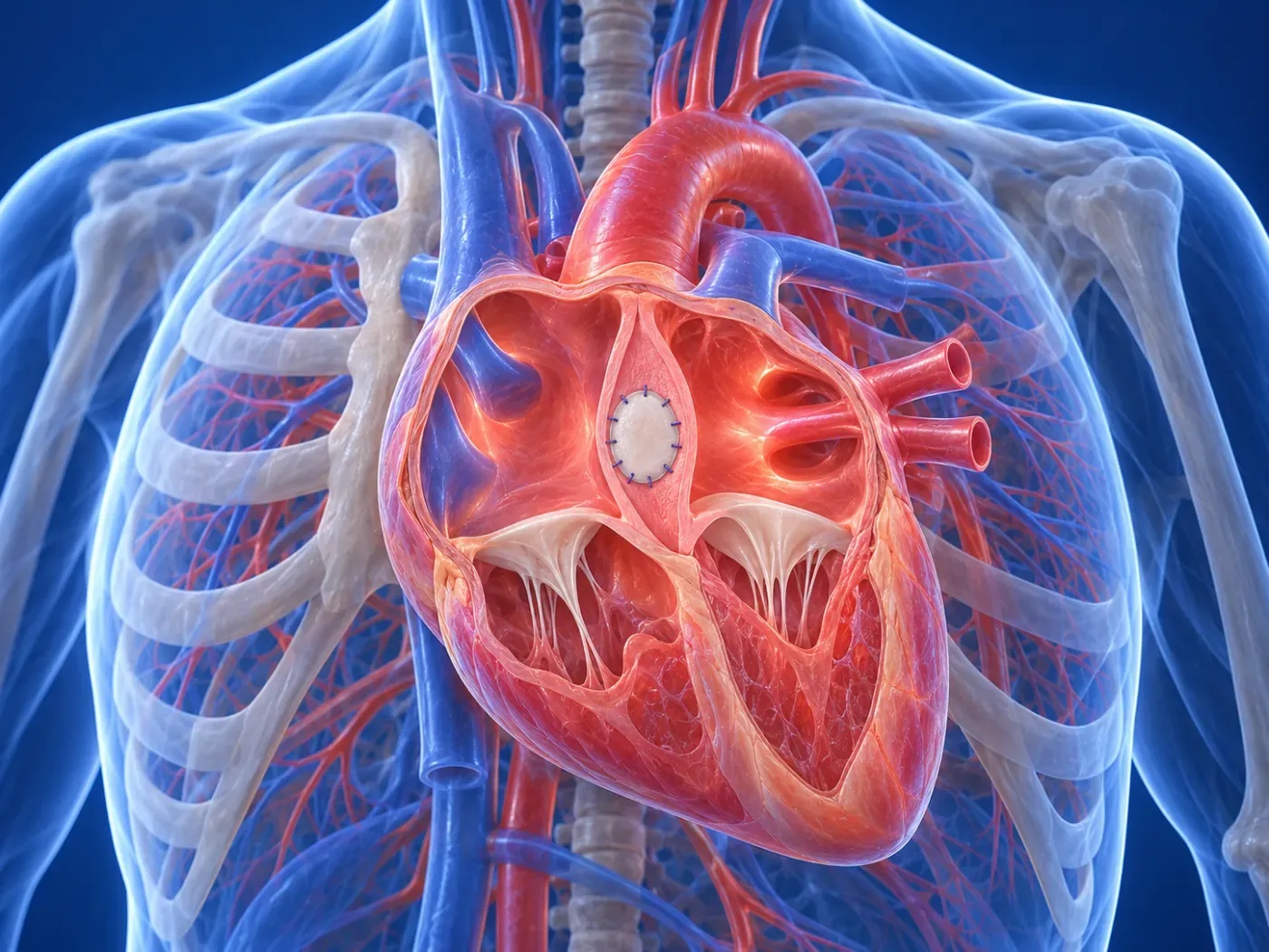
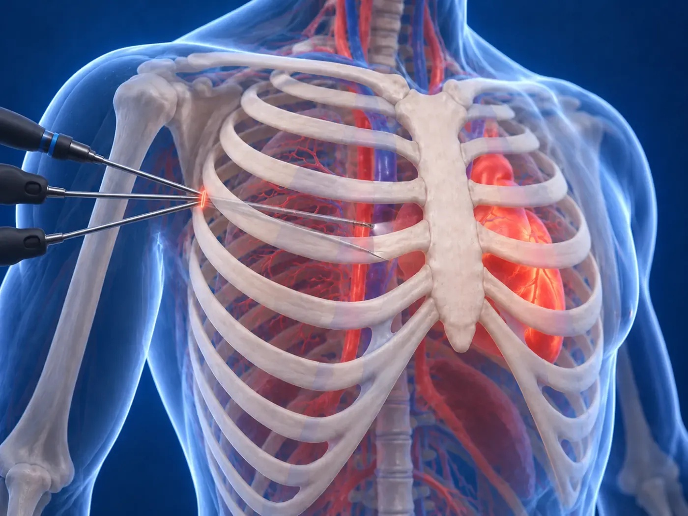
Coronary artery disease is caused by atherosclerosis narrowing or blocking the vessels feeding the heart muscle. In modern cardiac surgery, minimally invasive and endoscopic coronary artery bypass (MIDCAB) is performed on the beating heart through a small small incision under the left breast, completely without cutting the breastbone or connecting to a heart-lung machine.
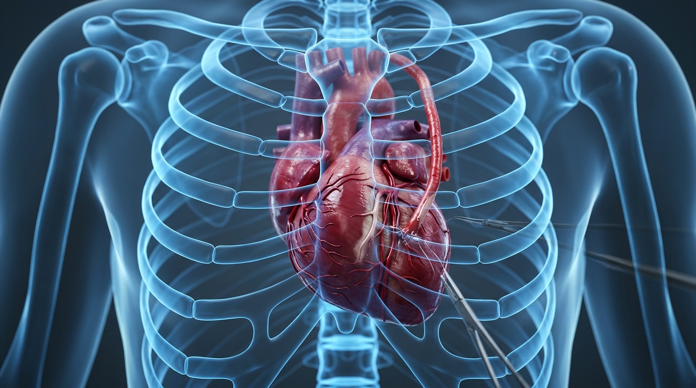
Coronary artery disease is caused by atherosclerosis narrowing or blocking the vessels feeding the heart muscle. In modern cardiac surgery, minimally invasive and endoscopic coronary artery bypass (MIDCAB) is performed on the beating heart through a small small incision under the left breast, completely without cutting the breastbone or connecting to a heart-lung machine.
Coronary Artery Disease and the Philosophy of Endoscopic Bypass
In traditional coronary bypass operations, the breastbone (sternum) is fully divided, and the patient is placed on a heart-lung cardiopulmonary bypass machine. This introduces significant risks of bleeding, systemic inflammation, neurocognitive dysfunction, and sternal bone dehiscence or infection, particularly in diabetic and elderly patients.
Dr. Taha Özkara utilizes high-definition endoscopic camera guidance and advanced coronary stabilization systems to access the left anterior descending (LAD) and diagonal coronary arteries through an intercostal small incision, ensuring maximum revascularization while completely sparing the thoracic skeleton.
Surgical Technique: LIMA Grafting & Beating Heart Microsurgical Precision
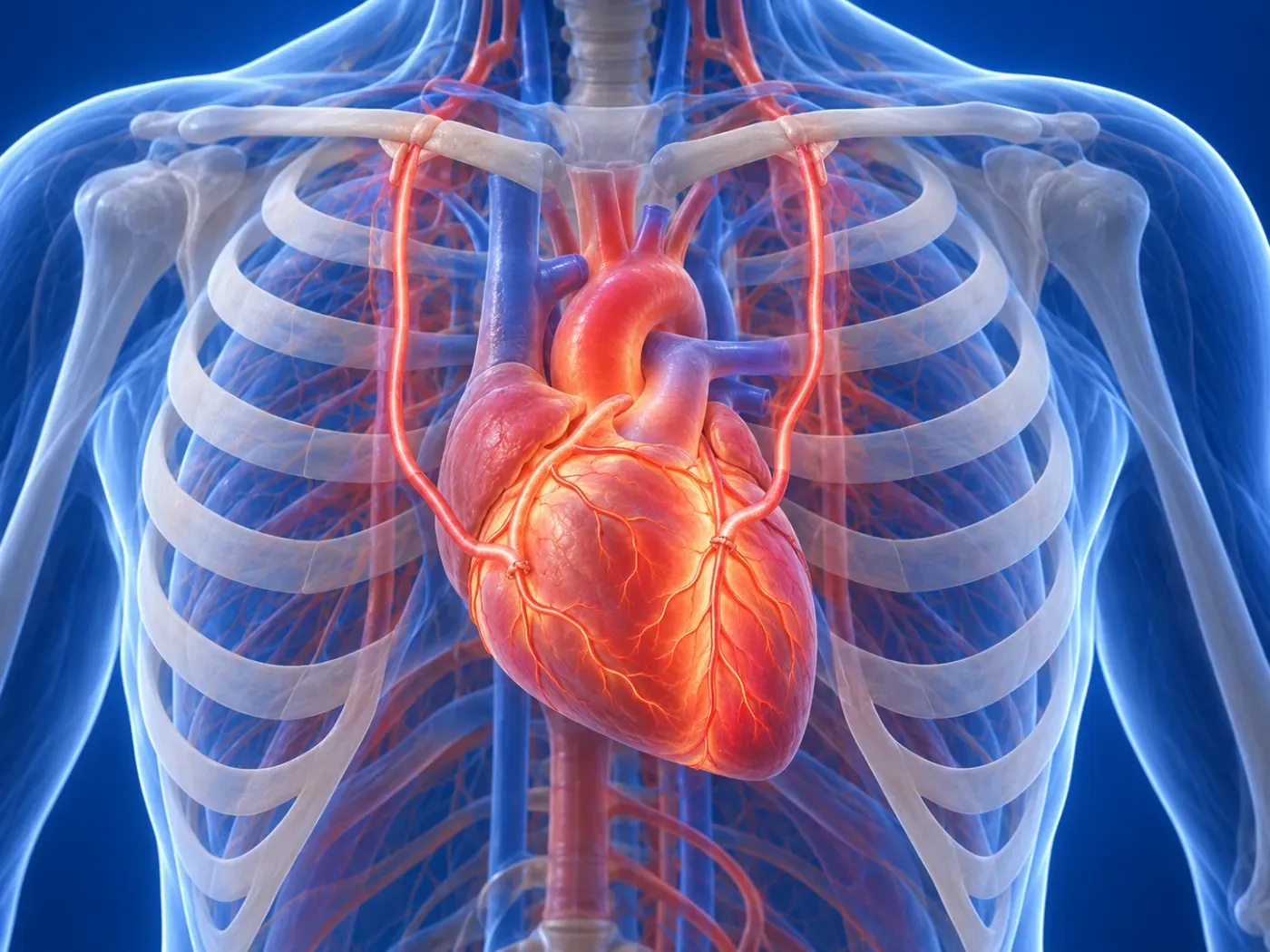
The Left Internal Mammary Artery (LIMA)—the gold standard coronary bypass graft with over 95% 10-year patency—is harvested under direct high-magnification endoscopic visualization through the small intercostal window. Special micro-vascular instruments allow delicate skeletonized or pedicled LIMA harvesting without any rib fracture.
Using specialized cardiac vacuum stabilizers, only the target diseased coronary segment is immobilized while the remainder of the heart continues beating normally. Under 4x-6x surgical loupes, a continuous microvascular anastomosis is constructed using 7-0 or 8-0 monofilament sutures, establishing immediate robust coronary myocardial perfusion.
Clinical Superiority & Advantages Over Traditional Open Bypass
By completely eliminating sternotomy and extracorporeal circulation, off-pump endoscopic bypass dramatically reduces systemic surgical trauma and accelerates patient recovery:
- The breastbone (sternum) remains completely intact; thoracic structural integrity is 100% preserved.
- Operated on the beating heart without cardiopulmonary bypass, drastically lowering stroke and renal risks.
- Minimal blood loss; blood transfusion requirement is virtually zero.
- No requirement for rigid chest corsets or sleeping flat on the back.
- Intensive care stay is typically under 24 hours, with hospital discharge in 3-4 days.
- Patients can return to driving, active work, and sports in just 10 to 14 days.
Recovery Period, Hospital Discharge, and Patient Suitability
Following surgery, patients are extubated within a few hours and transition from the intensive care unit to a private room the next morning. Because bone healing is not required, deep breathing exercises are pain-free and pulmonary complications are virtually eliminated.
Endoscopic coronary bypass is particularly ideal for patients with isolated LAD or multi-vessel disease suitable for hybrid revascularization, as well as high-risk diabetic, COPD, and elderly patients where traditional sternotomy carries elevated morbidities. Contact us with your coronary angiography images for a comprehensive evaluation.
FREQUENTLY ASKED
Frequently Asked Questions
Expert answers to the most common questions regarding minimally invasive heart surgery, recovery timelines, and surgical procedures.
TREATMENT TIMELINE
Your 4-Step Surgical Journey
Tetkiklerinizin incelenmesinden ameliyat sonrası aktif yaşama dönüşünüze kadar geçen 4 aşamalı konforlu ve güvenli cerrahi yolculuğunuz.
Preliminary Review
Comprehensive analysis of your Echo, Angiography, and CT scans.
Tailored Plan
Individual surgical strategy without sternotomy.
Endoscopic Surgery
High-precision closed operation with 3D camera guidance.
Swift Recovery
Discharge in 3-4 days and fast return to normal life.
OUR TREATMENT AREAS
Minimally Invasive and Endoscopic Heart Surgery
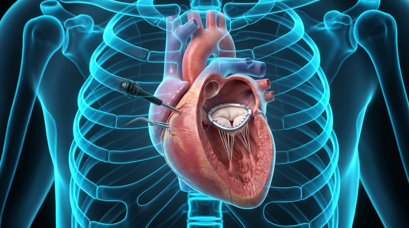
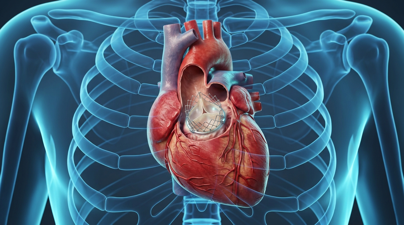
Innovative cardiovascular surgical procedures performed through small incisions with high-definition endoscopic camera guidance without cutting the breastbone.
EXPERT OPINION & APPOINTMENT
Schedule a Medical Consultation
Book your appointment for minimally invasive heart surgery, valve repair, and second opinion consultation.